

Published Oct 02, 2026 | 7:00 AM ⚊ Updated Oct 02, 2026 | 7:00 AM

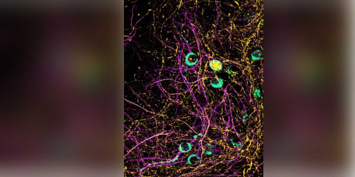
SARS-CoV-2 infected neurons in magenta, viral RNA in teal and α-Synuclein aggregates in yellow (Supplied)

Synopsis: Hyderabad researchers have found that viral RNA can push a Parkinson’s-linked brain protein towards harmful aggregation in neurons. Studying H1N1 and SARS-CoV-2 RNA, the team also identified DDX39A. This cellular enzyme can unwind these RNA structures and slow protein aggregation, offering clues to how viral infections may influence long-term Parkinson’s disease risk.
Every influenza and COVID infection sparks a fight inside the human body. Cells use their proteins to try to stop the virus from copying itself. Researchers in Hyderabad now report that this fight may leave something behind.
A team at Hyderabad’s CSIR-Centre for Cellular and Molecular Biology (CCMB) found that folded pieces of viral genetic material push a brain protein to stick together in clumps. These clumps mark Parkinson’s disease, a brain ailment that affects movement.
The team also found a defender protein that unravels the viral material, slowing the virus and the clumping. But the defender and the brain protein also join forces, and the authors said that the “teamwork” may carry a price. They described the result as “a mechanistic link between viral infections and an increased long-term risk” of Parkinson’s disease.
The work from Dr Swasti Raychaudhuri’s lab has appeared in Cell Reports. Scientists from IIT Kharagpur and the Academy of Scientific and Innovative Research were part of the team.
The results came from cells, mouse neurons and test tubes. Nobody has shown that flu or COVID gives a person Parkinson’s disease.
Also Read: How a refined-carb-heavy diet can raise metabolic and heart risks
Doctors noticed Parkinson-like illness after the 1918 flu pandemic. A recent analysis of health records from Finland and the UK Biobank tied H1N1 flu to a higher risk of Parkinson’s disease later in life.
Those records showed a link. They did not show how an infection could lead to a brain disease.
The CCMB researchers said the link has gained weight in recent years. “Importantly, viral infections have recently been recognised as a long-term risk factor for [Parkinson’s disease],” they noted.
Their study looked for the missing step, starting with the virus’ genetic material.
Also Read: The things we blame on age
Flu and COVID viruses store their instructions in Ribonucleic acid (RNA), a polymeric molecule that works like a recipe card inside cells. Human genes sit in DNA. These viruses use RNA instead.
Aanchal (left) and Swasti Raychaudhuri (right)
RNA does not always appear flat like a thread. The authors noted that “many RNA-virus genomes have complex secondary structures”. In plain terms, the RNA folds itself into knots.
Viruses use these knots to help them copy themselves. The team probed what happened when the knots met a protein called alpha-synuclein, which lives in neurons, the cells that carry signals in the brain.
In Parkinson’s disease, alpha-synuclein clumps together into amyloids. The clumps block signals between neurons and harm the cells.
The team first added RNA from ordinary cells to neurons and other cells that make the brain protein. Clumping sped up. DNA did not have the same effect.
The authors said the result pointed to the RNA’s folded shape, “implicating RNA secondary structures in this process”. The knots, and not just the extra genetic material, seemed to matter.
The team then turned to virus RNA. Mouse neurons received genetic material from an H1N1 flu virus. Within four days, clumps of the brain protein appeared around the nucleus, the control centre of the cell.
The researchers scanned the flu genome and tested each knot. One knot had the most effect. When the team broke its shape, the effect faded.
Knots from the COVID and the Japanese encephalitis viruses also sped up clumping, though with less force than the flu knot.
Also Read: Fake drugs in the market?
Cells do not remain still during an infection. The team studied a defender protein called DDX39A. It normally stays in the nucleus, but during some infections it moves out and grabs viral RNA.
DDX39A works like a zip puller. It unwinds RNA knots, and it needs ATP, the fuel that powers cells, to do the job.
In test-tube experiments, the flu knot made the brain protein gather into about 250 droplets per field of view. Samples without the knot showed four to five. Droplets are the first step towards clumps. When DDX39A and ATP joined the mix, the count fell to six or seven.
The unwinding hurts the virus too.
“The virus fails to replicate with its RNA structures dismantled, and thus, the viral load in the cells decreases,” Aanchal Jain, lead author of the study, explained. “At the same time, the unwinding of viral RNA’s secondary structure effectively slows down α-Synuclein amyloid formation.”
Put simply, one action gives the cell two wins. The virus copies itself less, and the brain protein clumps more slowly.
Also Read: Police uncover fake-medicine racket supplying life-saving drugs
RNA pieces made in a lab do not match a real infection. The authors said adding RNA knots directly “bypasses key features of natural viral infection”.
The team therefore infected neurons from mice bred to carry the human receptor that the COVID virus uses to enter cells.
After infection, the neurons built clumps of the brain protein around their nuclei. They also showed more of a chemical tag that marks the protein when it starts to clump.
Extra DDX39A in these neurons cut the clumps. It also lowered the amount of virus in the cells. A version of DDX39A that could not grab RNA failed to help.
Also Read: Young-onset Parkinson’s disease rising in India
The study came across a twist. The brain protein doesn’t just fight viral RNA. It also helps the defender.
The two proteins gather in droplets together, a process the team called co-condensation. Inside these droplets, DDX39A unwound RNA knots with about 50 per cent more activity than it showed alone.
That boost helps the cell fight the virus. But the authors noted that “co-condensation may initially serve a protective function, with large amyloid formation emerging as a long-term consequence.”
Order matters as well. When DDX39A reached the knots first, unwinding rose. When the brain protein reached the knots first, DDX39A failed to unwind them.
Dr Raychaudhuri said the outcome depended on which side gains ground.
“Even if there are protective mechanisms in the cells to prevent viral infections and avoid amyloid formation, there are situations that favour one kind of reaction more than the other,” he said. “These decide the outcomes in cells, and sometimes amyloid formation is accelerated in virus-infected cells.”
Amyloid formation is the clumping. The team described the whole picture as a three-way balance between the brain protein, the virus knots, and the defender. An infection adds viral RNA and shifts the balance.
The researchers did not say that flu or COVID gave anyone Parkinson’s disease. Dr Raychaudhuri stressed that most infections left no such mark.
“So, while not every viral infection leads to increased amyloid formation and neurodegenerative disease, repeated exposure might tilt the odds against us,” he said.
That line described a hypothesis, not a result. The study did not follow people or measure risk over time.
For COVID, the paper relied on a separate recent study. In that work, the COVID virus raised the clump-marking tag in human neurons and in a mouse model. That is a lab result and not proof of higher risk in people.
The experiments were run in cell lines, mouse neurons and test tubes. No experiment infected a live animal, and no person took part.
Most cell work also used seeds of pre-formed clumps to start the process. Those seeds speed up an event that could take far longer in a living brain.
A gap also existed between the lungs and the brain. The authors wrote that “both influenza and SARS-CoV-2 predominantly infect the respiratory tract”, and that how clumping might reach the brain “remains an open question.”
They pointed to reports that clumped protein placed in the gut or kidney could travel to the brain. Infection outside the brain, followed by travel of clumps inwards, “cannot be ruled out,” they noted. It remains a possibility, not a finding.
The authors said further work must remove DDX39A from cells, test a version that cannot unwind RNA, study how the proteins bind each other and infect animals.
Dr Raychaudhuri’s lab is now working to uncover the finer details of how a routine viral infection might quietly set the stage for Parkinson’s disease years later.
The authors named one direction. “Cooperative phase separation of DDX39A and α-Synuclein to mitigate viral infections is an intriguing possibility to investigate in vivo,” they wrote.
In vivo means performed or taking place in a living organism.
If more DDX39A helps cells clear the virus and slows clumping, a boost could help. If it also draws the brain protein into droplets, a boost could backfire. Only more experiments can show which effect wins.
For now, the study gives the field a map. It shows how viral RNA, a protein that fights viruses and a protein tied to Parkinson’s disease may push and pull on each other inside a cell.
(Edited by Majnu Babu).



